Client: Ali Tahmasebi, MD
Field: Ophthalmology / ocular oncology
Figure type: Two-panel clinical and pathology illustration
Used for: Manuscript
Delivered: High-resolution PNG and editable Adobe Illustrator file
The brief
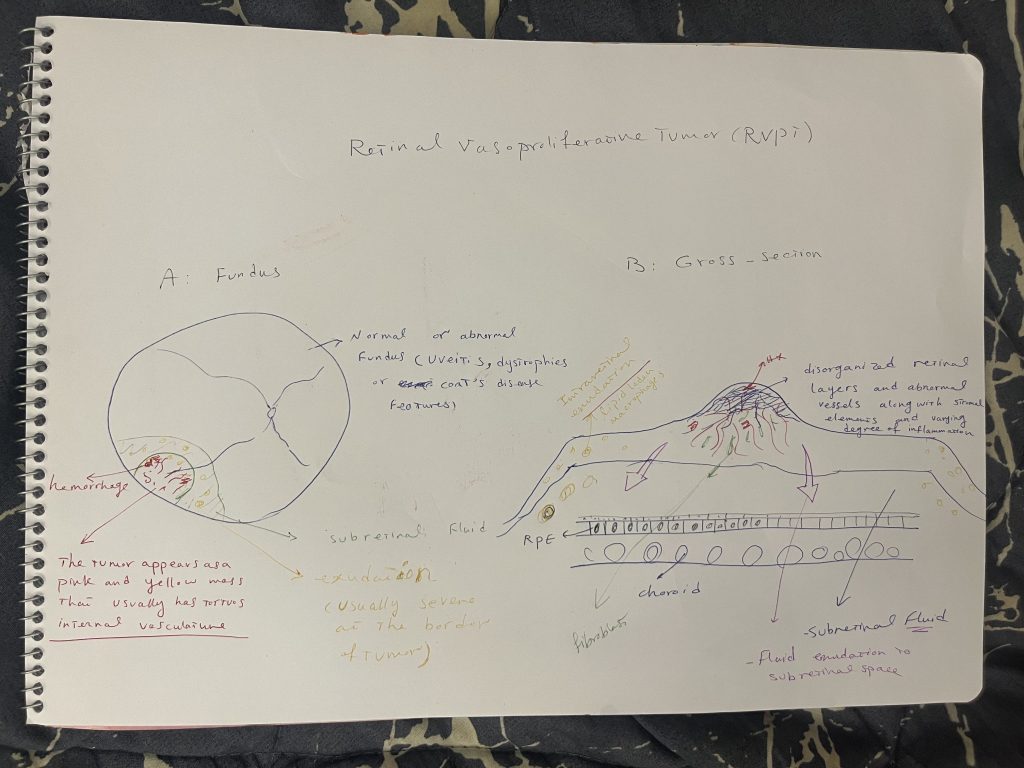
The client needed a figure explaining retinal vasoproliferative tumor, a rare benign vascular tumor of the retina. It had to show both how the tumor looks on examination and what its structure is beneath the surface. The starting point was a hand-drawn sketch.
The challenge
Ophthalmologists recognize RVPT by its appearance on fundoscopy. Its consequences, though, come from what happens inside the retina: exudation, fluid, and disorganized tissue. A single view could only tell half the story.
My approach
I split the figure into two matching panels.
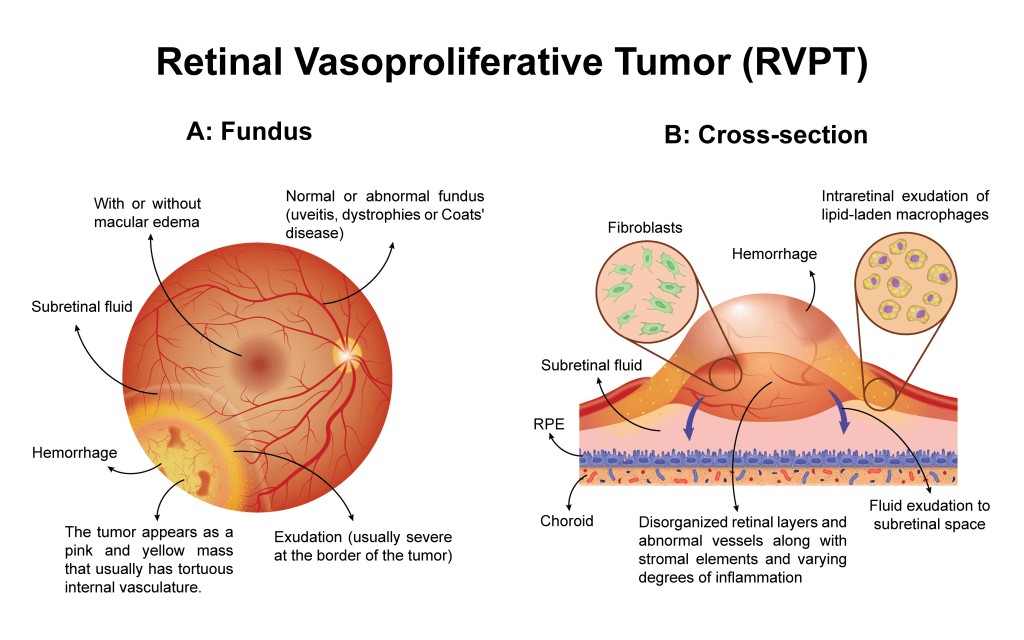
Panel A, Fundus: the view a clinician sees, with the tumor as a pink-yellow mass with tortuous internal vessels, surrounding exudation, subretinal fluid, and hemorrhage. A note on the associated conditions (uveitis, retinal dystrophies, Coats’ disease) gives clinical context.
Panel B, Cross-section: the same lesion in profile, showing the tumor’s position relative to the RPE and choroid, fluid leaking into the subretinal space, and disorganized retinal layers. Two zoom-in circles reveal the cellular components: fibroblasts and lipid-laden macrophages.
I used the same warm retinal palette in both panels, so the eye moves naturally from the clinical picture to the pathology behind it.
The result
A figure that connects bedside findings to tissue pathology, useful for a case report, a review, or teaching residents how to read what they see on fundoscopy.
Need a clinical or pathology figure? Start a project →