Client: Sana Hamidi, medical intern and researcher
Field: Neuro-oncology / cellular immunotherapy
Figure type: Central illustration for a systematic review
Published: 22 June 2026, Journal of Translational Medicine (Springer Nature)
Paper: LAK to CIK continuum in glioma immunotherapy: a systematic review of efficacy and safety outcomes
Delivered: High-resolution PNG and editable Adobe Illustrator file
The brief
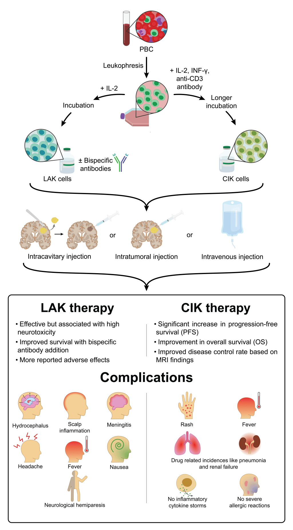
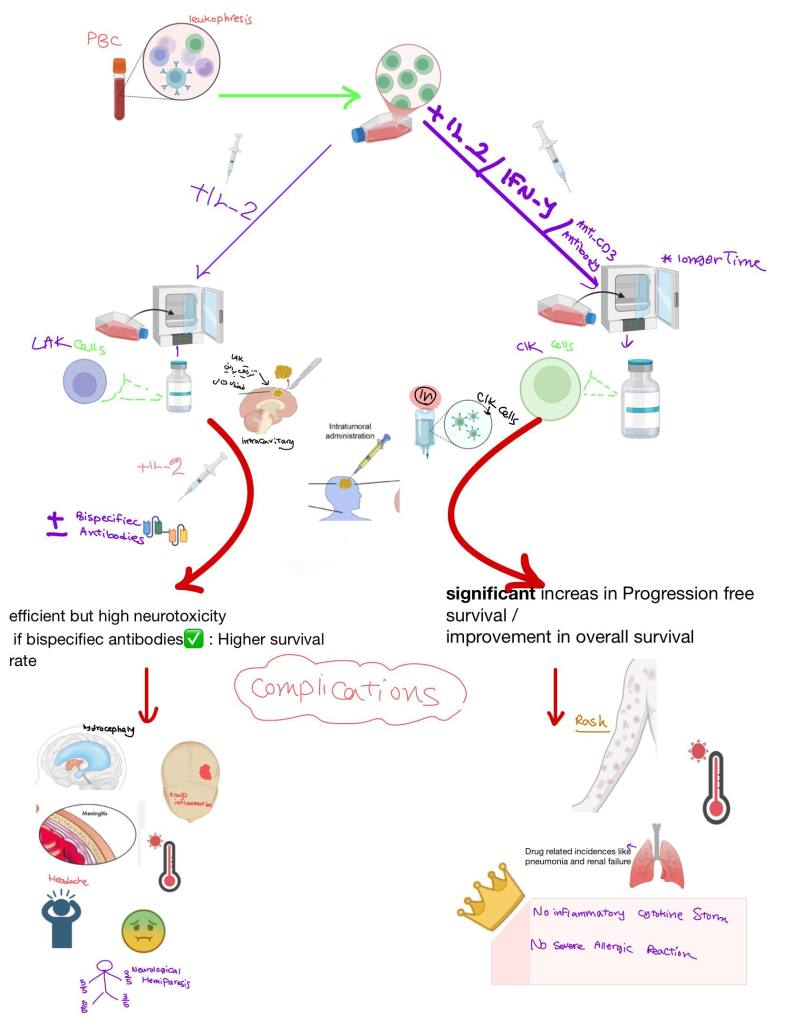
Sana’s systematic review compared two generations of adoptive cell therapy for glioma: lymphokine-activated killer (LAK) cells and cytokine-induced killer (CIK) cells. She needed one central figure that summarized the whole review: how each cell type is made, how it reaches the tumor, and how the two compare on efficacy and safety.
The challenge
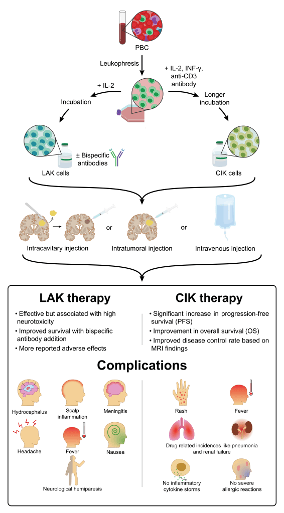
A systematic review covers a lot of ground. This figure had to carry three different kinds of information in one vertical flow: a manufacturing process (peripheral blood, leukapheresis, and culture with different cytokines and incubation times), three delivery routes (intracavitary, intratumoral, and intravenous), and the clinical outcomes and complications of each therapy.
My approach
I organized the figure as a single top-to-bottom story that branches and then rejoins.
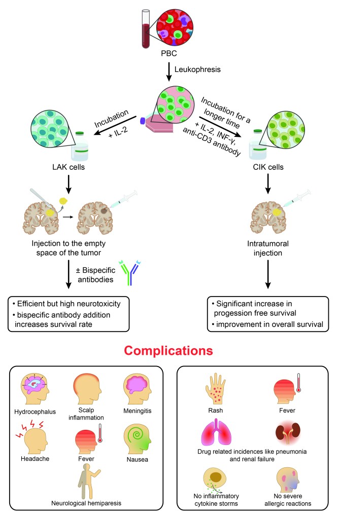
The top section starts from one blood sample and splits into two branches. LAK cells come from incubation with IL-2, optionally combined with bispecific antibodies. CIK cells come from IL-2, IFN-γ, and anti-CD3 antibody with longer incubation. Using the same vial and zoom-in style for both cell types makes the parallel easy to see.
The middle section brings the two branches back together into three delivery routes, drawn on matching brain sections so the routes can be compared directly.
The bottom section compares the therapies in two columns. Efficacy points sit above a grid of simple, recognizable complication icons, which readers can scan much faster than a table.
From draft to final
The project went through several rounds of edits and back-and-forth. Sana’s feedback after the first draft pushed the figure toward clarity: fewer words, a clearer flow, and a cleaner split between LAK and CIK outcomes.
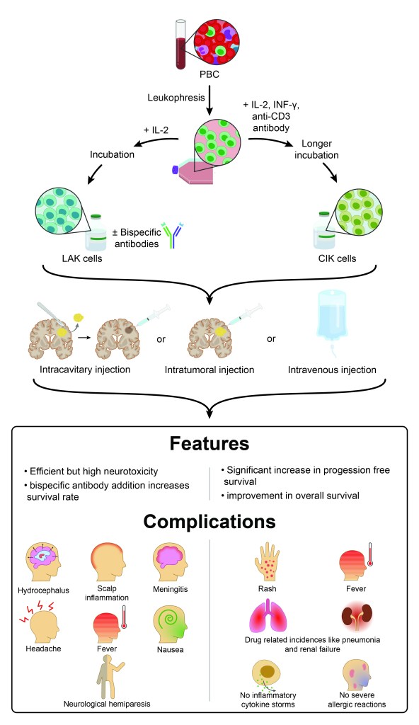
A final round of small adjustments produced the version that appears in the published paper.
The result
The figure was published as the central illustration of the review in the Journal of Translational Medicine. It condenses the paper’s main findings into one image that readers see before they read a word.
Writing a review or paper that needs a central figure? Start a project →