Accurate figures for glioma and brain tumour research: tumour microenvironment, immunotherapy, invasion and surgical anatomy. For labs, biotech teams and agencies working in neuro-oncology.
What I make
- Graphical abstracts and central illustrations for glioma papers
- Mechanism-of-action figures for cell therapies, vaccines, oncolytic viruses and small molecules
- Poster and deck figures for SNO, SITC, ESMO and AACR
- Surgical and anatomical views: resection cavity, tumour margins, mapping and delivery routes
- Journal cover art for accepted papers
Selected work
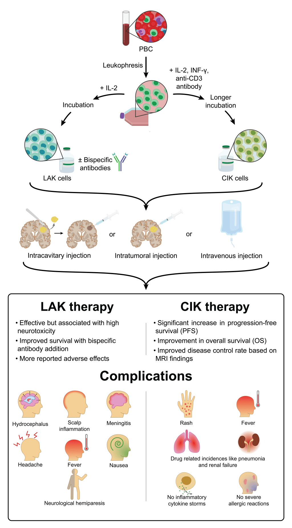
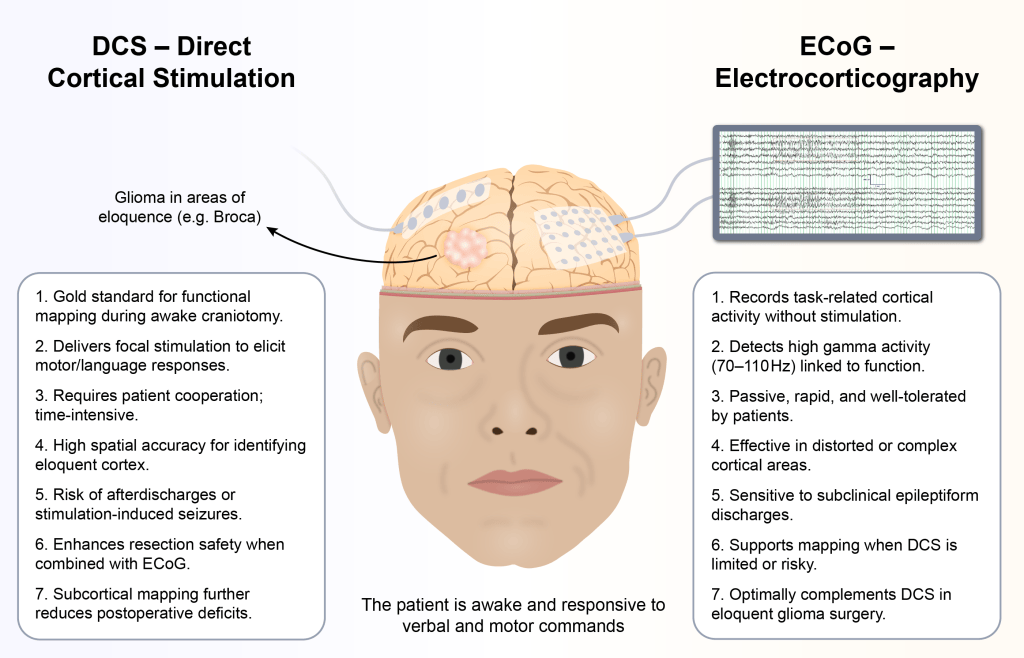
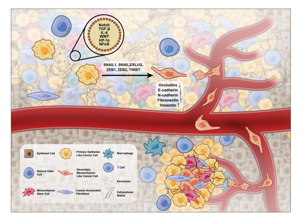
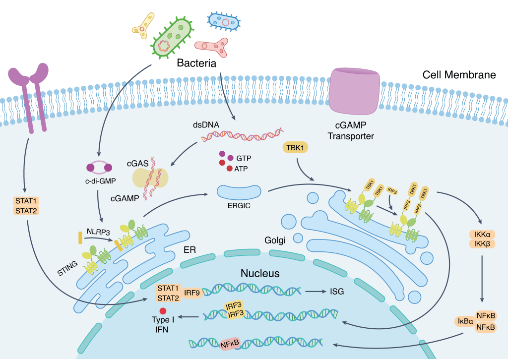
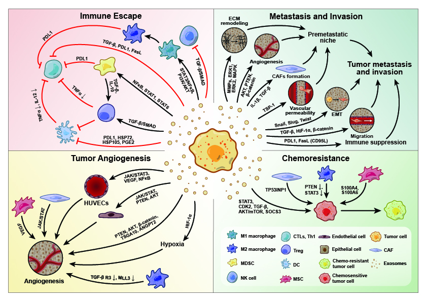
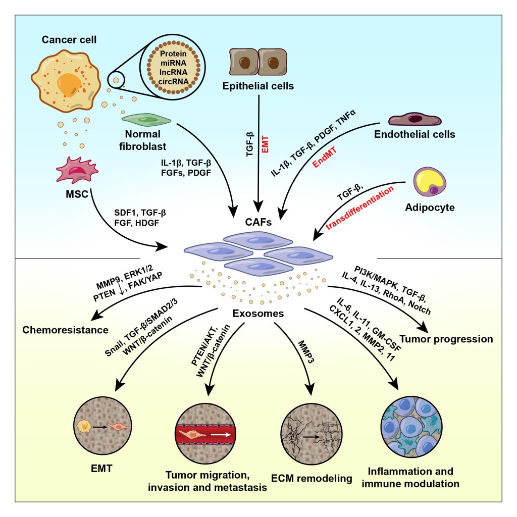
Why work with a medical researcher
I’m a medical intern and researcher, so I read the paper before I draw it. That means fewer rounds of corrections, mechanisms that are drawn the right way round, and figures that hold up in peer review. My figures are published in several Q1 journals, and every file comes editable and formatted to the journal’s specs.

