Client: Aysa Rezabakhsh, PhD
Field: Immunology / innate immune signaling
Figure type: Cell-signaling pathway illustration
Used for: Manuscript
Delivered: High-resolution PNG and editable Adobe Illustrator file
The brief
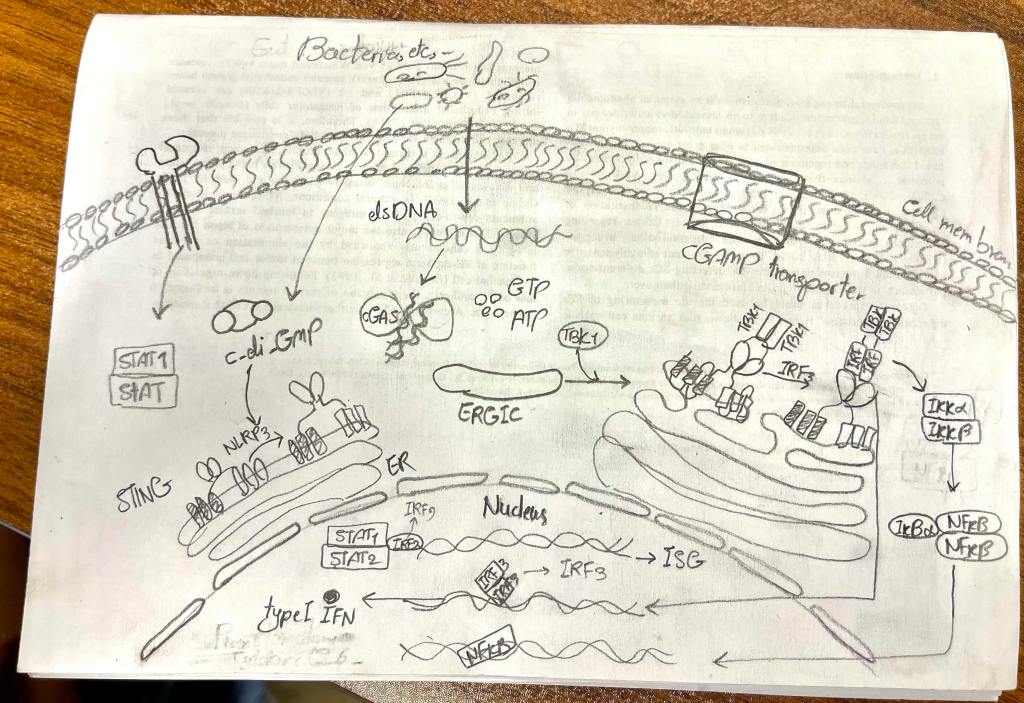
The researcher needed one figure explaining the cGAS–STING pathway, the system cells use to detect foreign or misplaced DNA in the cytoplasm and switch on an innate immune response. They sent a hand sketch of the pathway and the signaling molecules they wanted included.
The challenge
cGAS–STING signaling doesn’t happen in one place. It starts at the cell membrane, moves through the cytoplasm, travels from the endoplasmic reticulum through the ERGIC to the Golgi, and ends in the nucleus. On top of that, three signaling branches run in parallel: IRF3, NF-κB, and the JAK–STAT interferon loop. The figure had to keep every step in the right compartment while still reading as one connected story.
My approach
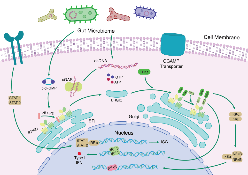
I drew the whole cell as a single stage, organized from the outside in, so the reader’s eye follows the signal the same way the cell does.
Outside the cell: bacteria at the top release dsDNA and the bacterial second messenger c-di-GMP. A cGAMP transporter in the membrane hints at signaling between neighboring cells.
In the cytoplasm: cGAS binds the DNA and uses GTP and ATP to make its second messenger, which activates STING sitting on the endoplasmic reticulum.
Through the secretory pathway: STING moves from the ER via the ERGIC to the Golgi, where it recruits TBK1 and IRF3. Drawing the organelles at full size made this trafficking step, which is often skipped in pathway diagrams, easy to follow.
In the nucleus: three readouts sit on their own DNA strands: IRF3 driving type I interferon, NF-κB released by the IKK complex, and STAT1, STAT2 and IRF9 inducing interferon-stimulated genes. Color-coded protein tags keep each branch distinct.
I changed the color for a more eye-friendly palette.
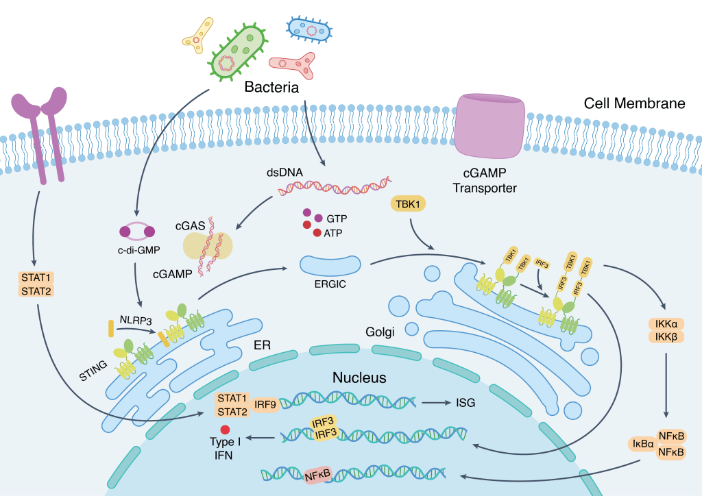
The result
A complete map of cGAS–STING signaling that shows not just which molecules are involved but where in the cell each step happens. It works as a review figure, a grant background figure, or a teaching slide on innate immunity.
Working on immune signaling or another multi-step pathway? Start a project →