Client: Dr. Reza RahbarGhazi’s group
Field: Cell biology / extracellular vesicles / cancer biology
Figure type: Pathway illustration with a molecular inset
Published: Cell Communication and Signaling (2024), Tumorigenic and tumoricidal properties of exosomes in cancers; a forward look (figure 1)
Delivered: High-resolution PNG and editable Adobe Illustrator file
The brief
This review on the dual role of exosomes in cancer needed an opening figure that explained the basics: how exosomes are made inside a cell and what they carry. I designed all six illustrations for the paper, and this one set the visual style for the rest.
The challenge
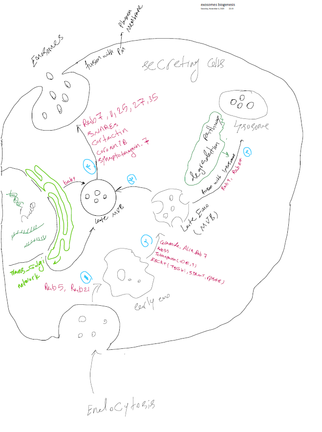
The figure had to do two jobs at different scales. At the cell level, it needed to show the full route from endocytosis to release, including the branch where vesicles are sent to the lysosome instead. At the molecular level, it needed to show the dozens of proteins, lipids, and nucleic acids that make up an exosome. Putting both in one view risked making either one unreadable.
My approach
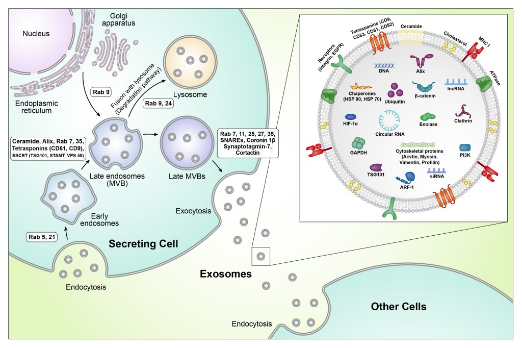
Left, the secreting cell: the pathway runs from endocytosis through early endosomes, late endosomes, and multivesicular bodies to exocytosis. The regulators of each step (Rab GTPases, ESCRT proteins, tetraspanins, SNAREs) sit in labeled boxes beside the step they control. A separate arrow shows the degradation route to the lysosome.
Right, a zoomed-in exosome: one released vesicle is enlarged into an inset. Its membrane shows tetraspanins, receptors, MHC I, ceramide, and cholesterol. Its interior shows the cargo, including DNA, lncRNA, siRNA, circular RNA, chaperones, signaling proteins, and cytoskeletal proteins. Each molecule has its own simple, consistent icon, so the inset reads like a labeled parts list.
Bottom right, the receiving cell: released exosomes are taken up by a neighboring cell, which completes the story from production to delivery.
The result
A figure that works at two scales at once: the pathway for readers who want the process, and the inset for readers who want the composition. It became the opening figure of a published review and set the style for the paper’s other five illustrations.
Working on extracellular vesicles or another cell-biology topic? Start a project →