Client: Dr. Reza RahbarGhazi’s group
Field: Cancer biology / metastasis
Figure type: Tissue-scene illustration
Published: Cell Communication and Signaling (2024), Tumorigenic and tumoricidal properties of exosomes in cancers; a forward look (figure 4)
Delivered: High-resolution PNG and editable Adobe Illustrator file
The brief
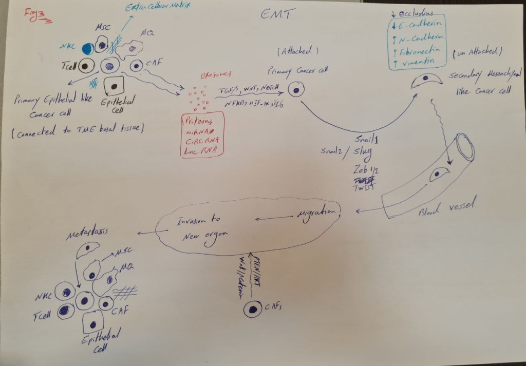
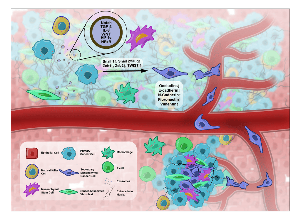
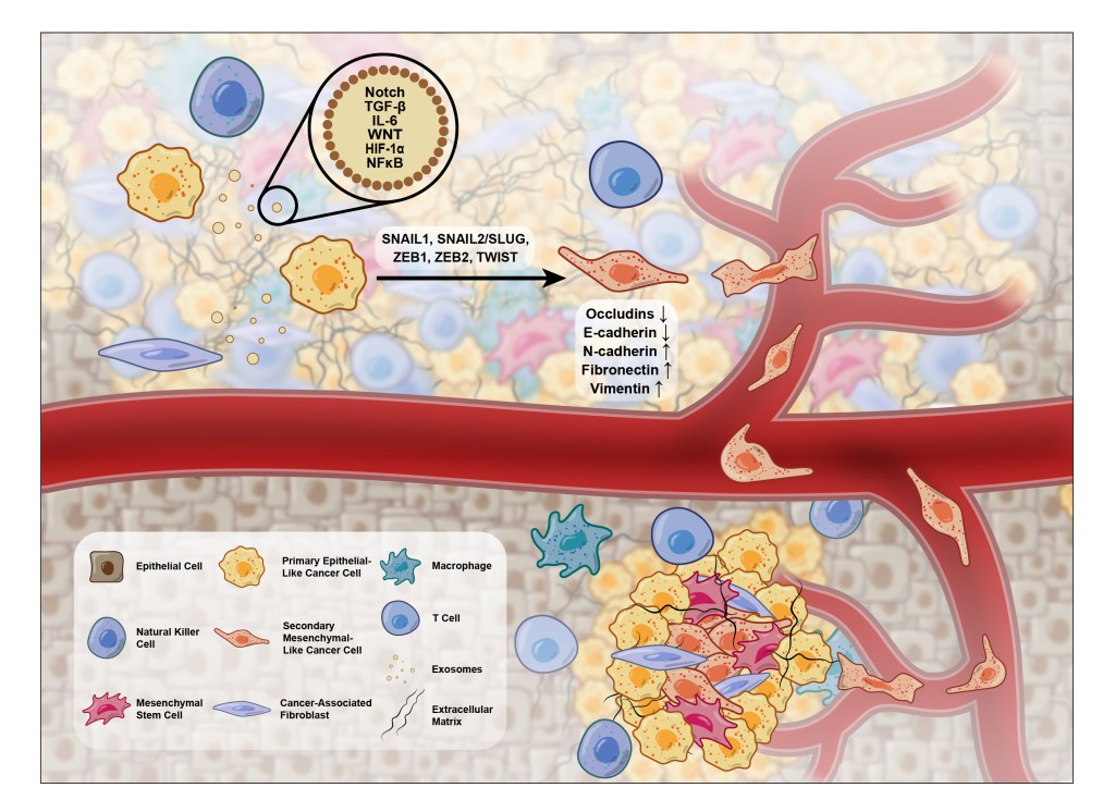
The fourth figure in this exosome review covered epithelial–mesenchymal transition (EMT), the change that lets cancer cells leave their original tumor and spread. The authors wanted it to feel like a real tissue rather than a diagram.
The challenge
EMT is a process that unfolds over space and time: a cell changes shape, loses its connections, and moves toward a vessel. A flat pathway diagram can list the molecules but cannot show the movement.
My approach
A scene instead of a diagram: I painted a crowded tumor microenvironment with immune cells, fibroblasts, stem cells, and extracellular matrix, all slightly blurred, so the cells taking part in EMT stand out in sharp focus.
The story moves left to right: a primary cancer cell receives exosome signals (Notch, TGF-β, IL-6, WNT, HIF-1α, NF-κB), switches on the EMT transcription factors, changes shape, and enters a blood vessel.
Marker changes beside the cell: falling occludins and E-cadherin and rising N-cadherin, fibronectin, and vimentin sit right where the transition happens.
A labeled legend: eight cell types, the exosomes and the extracellular matrix are labeled in a semi-transparent panel, so the busy scene stays readable.
The result
A figure that shows EMT as something happening in living tissue. It is one of my highest-rated pieces and a good example of a painterly style applied to a scientific figure.
Want a scene-style illustration for your paper or grant? Start a project →