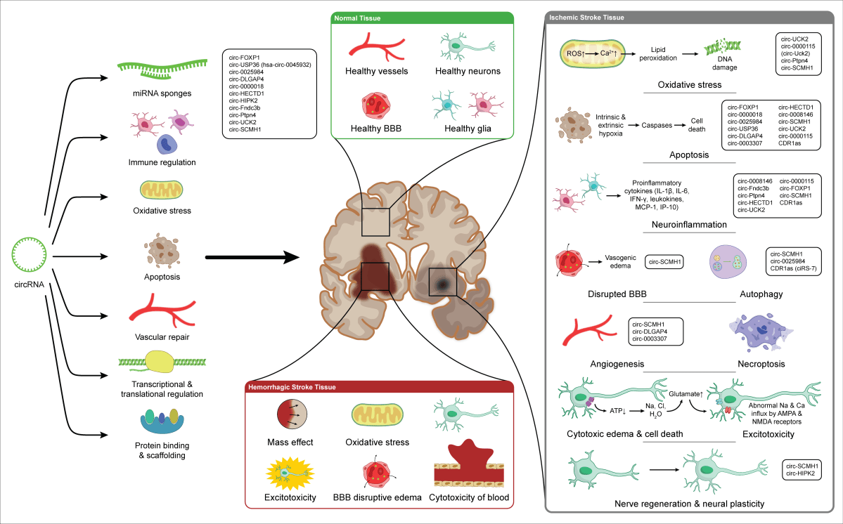
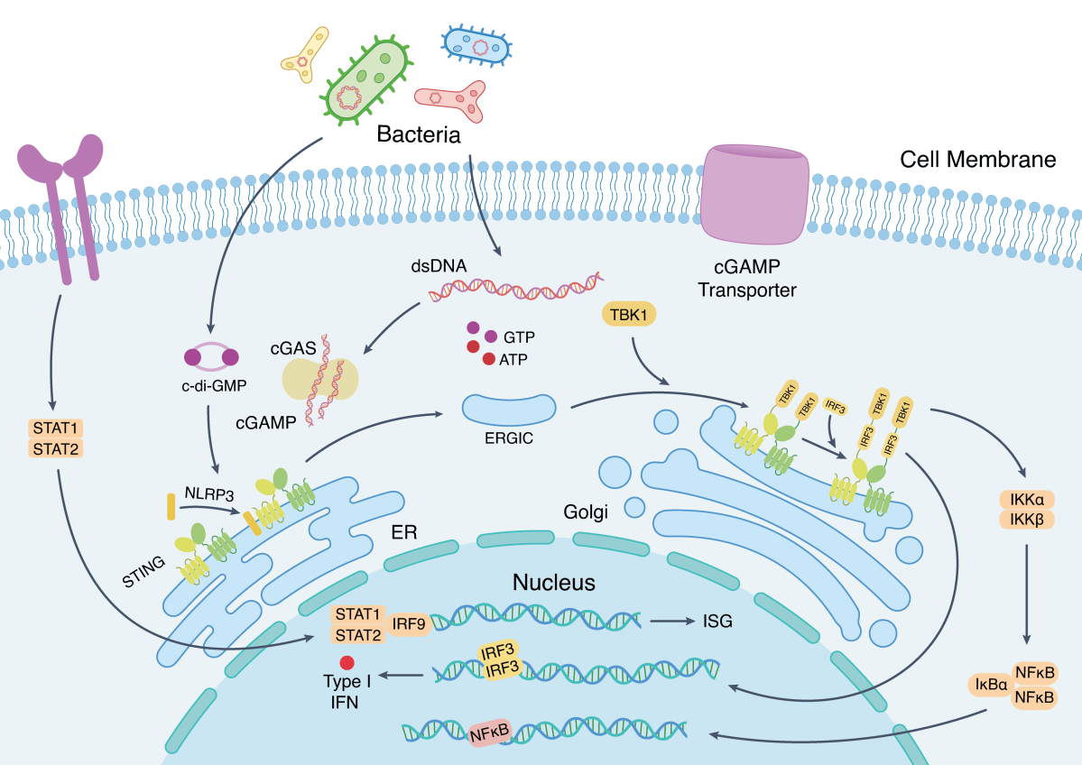
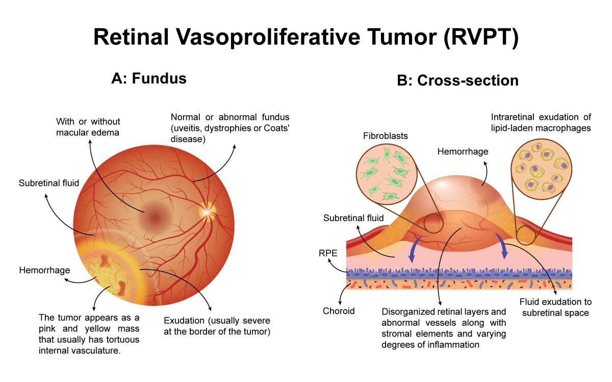
Client: Dr. Reza RahbarGhazi’s group
Field: Cancer biology / extracellular vesicle therapeutics
Figure type: Source-to-effect flow illustration
Published: Cell Communication and Signaling (2024), Tumorigenic and tumoricidal properties of exosomes in cancers; a forward look (figure 5)
Delivered: High-resolution PNG and editable Adobe Illustrator file
The brief
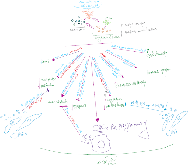
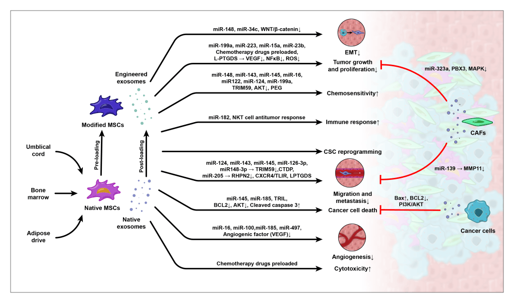
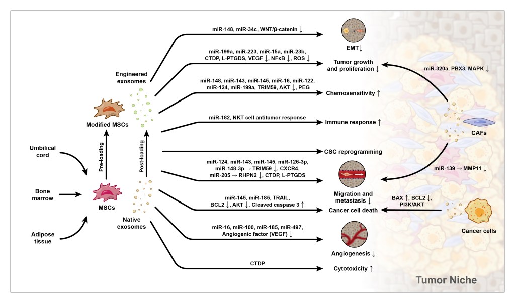
After several figures on how exosomes help tumors, the review turned to the other side: how exosomes from mesenchymal stem cells (MSCs) can be used against cancer. This figure needed to show where these exosomes come from, how they can be engineered, and what they do in the tumor.
The challenge
The figure had to carry a lot of molecular detail, with dozens of microRNAs and proteins, while staying a clear left-to-right story from source to effect.
My approach
Left, the source: MSCs from umbilical cord, bone marrow, and adipose tissue release native exosomes. A second route shows engineered exosomes, loaded either before release (pre-loading) or after (post-loading).
Middle, the cargo: each arrow carries the specific microRNAs and proteins behind its effect, so researchers can find the molecule they care about.
Right, the effects: nine outcomes line up in a single column, from reduced EMT and tumor growth to increased chemosensitivity and cancer cell death. The same tissue-window circles from the paper’s other figures make the link between figures obvious.
Background, the tumor niche: a faded tumor microenvironment with CAFs and cancer cells sits behind the effects, so the reader always knows where the action is.
The result
A dense figure that still reads as one clear flow, which makes it useful for both a quick overview and a detailed lookup of specific molecules.
Working on EV therapeutics or engineered exosomes? Start a project →