Client: Dr. Reza RahbarGhazi’s group
Field: Cell biology / extracellular vesicles
Figure type: Textbook pathway illustration
Published: Chapter in an Elsevier book
Delivered: High-resolution PNG and editable Adobe Illustrator file
The brief
This figure was made for a chapter in an Elsevier book on extracellular vesicles. Textbook figures have a different job from journal figures: they have to teach a process to students who may be seeing it for the first time.
The challenge
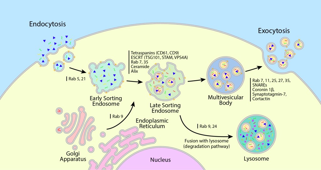
The endosomal pathway has several stages, each regulated by its own set of Rab GTPases and sorting proteins. For students, the figure needed to show the order of the stages clearly first, with the molecular detail as a second layer.
My approach
A single cell, one direction: cargo enters by endocytosis on the left and leaves as exosomes on the right, passing through early sorting endosomes, late sorting endosomes, and the multivesicular body along the way.
A clear second route: a separate arrow sends late endosomes to the lysosome, labeled as the degradation pathway, with the Golgi feeding in from below.
Regulators beside each arrow: the Rab proteins, tetraspanins, ESCRT components, and SNAREs are listed next to the step they control, marked with a simple bar instead of a box.
A flat, print-friendly style: solid colors and clean outlines reproduce well in print and stay readable at small sizes.
The result
A clean teaching figure that students can follow step by step, now part of a published Elsevier book.
Need figures for a textbook or teaching material? Start a project →