Client: Dr. Reza RahbarGhazi’s group
Field: Cell biology / extracellular vesicles
Figure type: Numbered mechanism illustration
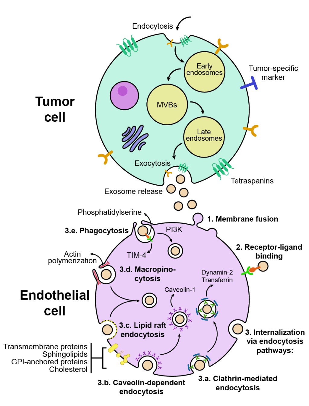
Published: Cancer Cell International (2024), Exosomes and breast cancer angiogenesis; highlights in intercellular communication (figure 3)
Delivered: High-resolution PNG and editable Adobe Illustrator file
The brief
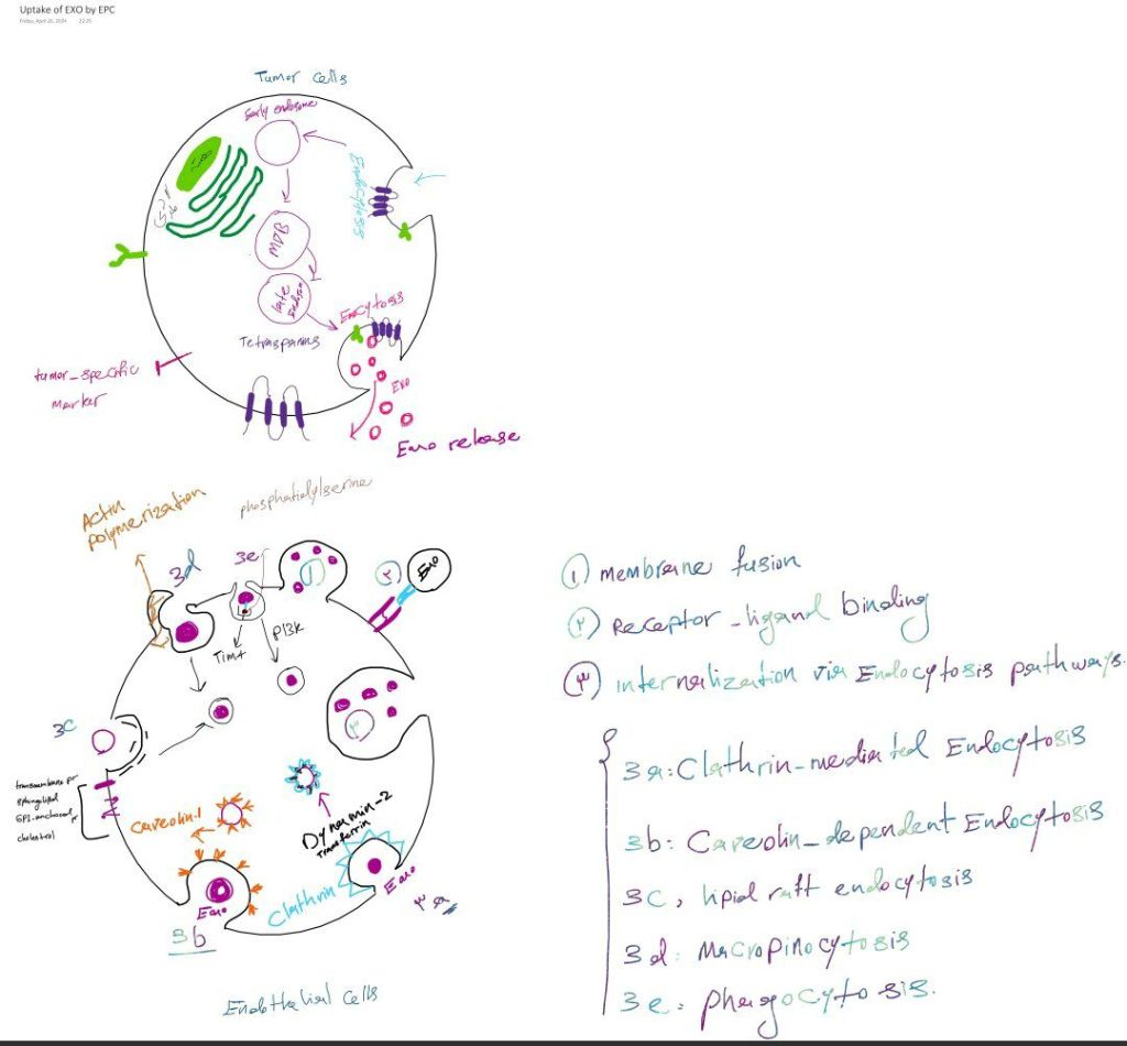
The last of my three figures for this breast cancer review explained how exosomes released by a tumor cell actually get into the endothelial cells they target.
The challenge
There are seven distinct uptake routes, and several of them look alike when drawn: a vesicle being pulled into a pocket of membrane. Each needed its own visual signature.
My approach
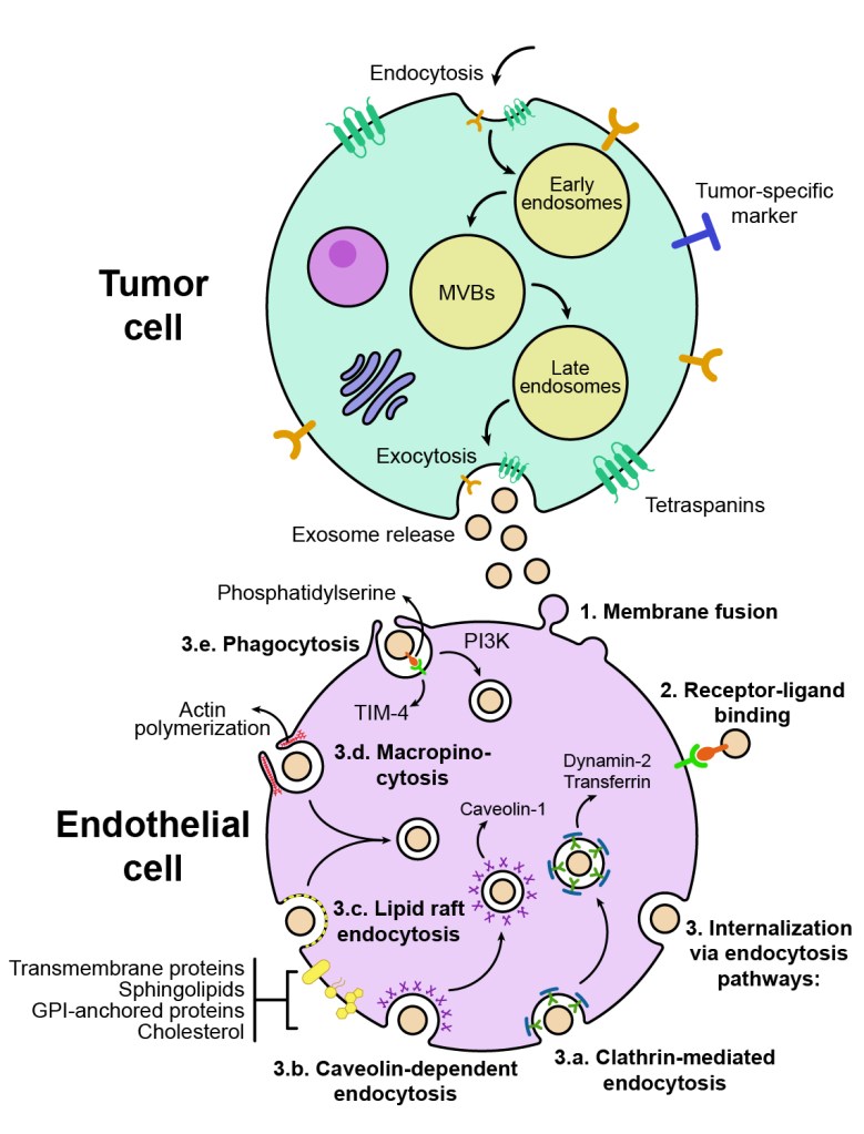
Sender above, receiver below: the tumor cell at the top releases exosomes downward, and the endothelial cell below takes them in, so the direction of travel is obvious.
Numbered routes: membrane fusion, receptor–ligand binding, and five endocytosis pathways (clathrin-mediated, caveolin-dependent, lipid raft, macropinocytosis, and phagocytosis) are numbered around the edge of the cell.
A visual signature for each route: clathrin as a blue-green coat, caveolin as purple crosses, lipid rafts as a dotted yellow membrane patch, macropinocytosis as an actin-driven ruffle. Each route can be recognized from its shape alone.
The result
A reference figure that lays out every uptake route in one place, useful for reviews, methods introductions, and teaching.
Need a mechanism figure for your paper? Start a project →